CHIARI MALFORMATIONS
WHAT ARE CHIARI MALFORMATIONS?
Chiari malformations (CMs) are structural defects in the cerebellum, the part of the brain that controls balance. Normally, the cerebellum and brainstem sit in a triangular space at the lower rear of the skull, where the brain is linked to the spinal canal. A Chiari malformation happens when part of the cerebellum is located below the foramen magnum, which connects the posterior fossa with the spinal canal.
Chiari malformations develop when the space inside the skull is smaller than normal, causing the cerebellum and brainstem to be pushed downwards towards the foramen magnum and the cervical spinal canal. The resulting pressure on the cerebellum and brainstem may affect functions controlled by these areas and block the flow to and from the brain of cerebrospinal fluid (CSF), a clear liquid that surrounds and protects the brain and spinal cord, to and from the brain.
CAUSES OF CHIARI MALFORMATIONS
The exact cause of Chiari malformations is yet unknown. It is believed that Chiari malformations are the result of a structural defect produced during the development of the foetus, but they may also be genetically-associated: a tendency to develop Chiari malformations has been seen to run in certain families. Other possible causes include exposure to harmful substances, deficiency of some vitamins or nutrients in the mother’s diet during foetal development, injury, infection, or aging.
WHAT ARE THE TYPES OF CHIARI MALFORMATIONS?
Chiari malformations are classified by the severity of the disorder and by the parts of the brain that are herniated into the cervical spinal canal.
Type I: these malformations arise from the herniation of the cerebellar tonsils, a structure in the lower part of the cerebellum, into the foramen magnum, blocking the flow of CSF towards the cervical spinal canal. The symptoms of this type of CM first appear in the adolescence or adulthood. Often, however, the CM is found by accident when performing a brain and neck MRI on investigating for another condition.
 |
|
Chiari malformation type I with syringomyelia
|
Type II: also called Arnold-Chiari malformations, these malformations involve the extension of the cerebellum and brainstem tissue through the foramen magnum. The cerebellar vermix, the nerve tissue that connects the two halves of the cerebellum, may be partially complete or absent. This type of CM is usually accompanied by a myelomeningocele, a form of spina bifida that occurs when the spine and spinal canal do not close before birth, causing the rear part of spinal canal and its protective membrane (duramater) not to close and so exposing the nervous tissue. A myelomeningocele usually results in partial or complete paralysis of the area below the spinal opening.
Type III: this is the most severe form of CM. The cerebellum and brainstem are herniated trough the foramen magnum and into the cervical spinal canal. The brain’s IVth ventricle, a cavity that links the upper parts of the brain and through which CSF flows, may also be herniated through the foramen magnum and into the spinal canal. In rare cases, the herniated cerebellar tissue may cause an occipital encephalocele, a bag located at the lower rear of the head containing CSF and nervous tissue that has been herniated through a small abnormal opening at the back of the skull. This type of CM causes severe neurological deficits.
Type IV: these malformations are the rarest and involve an incomplete or underdeveloped cerebellum, a condition known as cerebellar hypoplasia. The cerebellar tonsils are located further down the spinal canal than they normally would be, part of the cerebellum is herniated, and some parts of the skull and spinal canal may be visible.
Another form of the disorder, known as type 0, does not involve any protrusion of the cerebellum through the foramen magnum, but headache and other symptoms of CM are present. This type is, however, under debate by some scientists.
SYMPTOMS OF CHIARI MALFORMATIONS
Not all people with a Chiari malformation show symptoms, and often the malformation is discovered by chance on performing a brain and neck MRI that was carried out to investigate some other condition. Patients with symptomatic CM may complain of pain in the neck, balance problems, muscle weakness, numbness or other abnormal feelings in the arms or legs, vertigo, vision problems, difficulty swallowing, ringing or buzzing in the ears, hearing loss, vomiting, insomnia, depression, or headache that gets worse on coughing. The symptoms may differ from patient to patient depending on the build-up of CSF and the resulting pressure on nervous tissue.
In infants, symptoms may include difficulty swallowing, irritability when being fed, excessive drooling, weak crying, vomiting, weakness in the arms, stiff neck, difficulty to breathe, and inability to gain weight. Children may snore while asleep, and adolescents and adults with a CM that do not initially show symptoms may start to show them over the years.
ARE OTHER CONDITIONS ASSOCIATED WITH CHIARI MALFORMATIONS?
Individuals with a Chiari malformation may show the following conditions:
Hydrocephalus is an excessive build-up of CSF in the brain. A Chiari malformation can block the normal flow of CSF, increasing the pressure inside the head. This increase in pressure can cause mental defects as well as an enlarged or misshapen skull, and severe hydrocephalus can be fatal if left untreated. Hydrocephalus is most commonly associated with type II, but it can occur with any type of CM.
Spina bifida is the incomplete development of the spinal cord and/or its protective covering. The bones around the spinal canal do not form properly, leaving part of the spinal cord exposed and resulting in flaccid partial or complete paralysis. Patients with CM type II usually show a myelomeningocele, a form of spina bifida that occurs when some bones of the spine and the spinal cord coverings are not correctly formed and so part of the nervous tissue (a bag of nervous tissue and CSF) is herniated outwards though a hole.
Syringomyelia is a disorder in which a CSF-filled tubular cyst (a syrinx) forms inside the spinal cord. As it grows it destroys the tissue of the spinal cord core, resulting in pain, weakness, and stiffness in the back, shoulders, arms or legs. Other symptoms include headaches as well as loss of sensitivity to temperature, especially in the hands, and intense pain in the neck and arms.
Tethered cord syndrome occurs when the spinal cord attaches itself to the coverings that surround it. This progressive disorder happens due to the abnormal stretching of the spinal cord and can result in permanent damage to muscles and nerves in the lower trunk and legs. Children with a myelomeningocele have a high risk of developing a tethered cord syndrome later in life.
 |
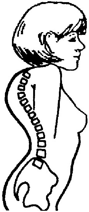 |
Spinal deviations are common amongst patients suffering from syringomyelia or a Chiari malformation type I. Two types of spinal curvatures are associated with Chiari malformations: scoliosis, a lateral bending of the spinal column to the left or to the right, and kyphosis, a forward bending of the spinal column. Spinal deviations are seen most often in children with Chiari malformations whose skeleton has not fully developed.
|
|
Scoliosis
|
Kyphosis
|
Chiari malformations may also be associated with certain hereditary syndromes characterised by neurological and skeletal abnormalities, as well as with other disorders that affect bone formation and growth, fusion of the segments of cervical vertebrae, and bony coverings in the brain.
HOW COMMON ARE CHIARI MALFORMATIONS?
In the past, it was estimated that Chiari malformations occurred in 1/1000 births. With the implementation of MRI, however, it has been shown that Chiari malformations are much more common; for, since the majority do not show symptoms, they were not detected until MRI appeared. Chiari malformations occur more often in women than in men and CMs type II are seen more often in certain groups, e.g. people of Celtic background.
HOW ARE CHIARI MALFORMATIONS DIAGNOSED?
Many people with Chiari malformations show no symptoms and their CMs are discovered by change during the radiological investigations to study another disorder. Once the condition is suspected, the doctor will perform a physical exam and will check the patient’s memory, cognition, balance (controlled by the cerebellum), reflexes, sensitivity, and motor skills (controlled by the spinal cord). The doctor may also order one of the following diagnostic tests:
Plain X-rays of the head and neck cannot reveal a Chari malformation but can identify bone abnormalities that are associated with Chiari malformations.
A computed tomography (CT scan) uses X-rays and a computer to produce 2D images of bones, blood vessels, some brain tumours, cysts and hydrocephalus. CT scans can, thus, show hydrocephalus as well as bone abnormalities associated with Chiari malformations.
 |
Magnetic Resonance Imaging (MRI) is the neuro-imaging procedure most often used to diagnose Chiari malformations. It uses radio waves and a powerful magnetic field to produce either a detailed 3D image or a 2D “slice” of body structures, including soft tissues, organs, bones and nerves.
|
|
MRI image of Chiari malformation type I
|
HOW ARE CHIARI MALFORMATIONS TREATED?
Some Chiari malformations are asymptomatic and do not interfere with the daily activities of the patient. In other cases, medication may alleviate certain symptoms, such as pain.
Nonetheless, surgery is the only treatment available which can correct functional disturbances or halt the progression of the damage to the central nervous system. Though sometimes more than one surgery is needed, most patients who have surgery feel a reduction in their symptoms and/or prolonged periods of relative stability.
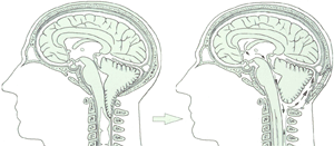
Posterior fossa decompression is a procedure carried out on adult CM patients to make more space for the cerebellum and to relieve the pressure on the cervical spinal cord. This procedure involves making an incision at the back of the head and removing a small portion of the base of the skull (and sometimes of the upper part of the cervical spinal cord) to correct the irregular bony structure. The neurosurgeon may, occasionally, partially remove the cerebellar tonsils. Since this surgical technique involves the partial destruction of nervous tissue it is usually not recommended.
 |
|
LEFT: Chiari malformation type I with syringomyelia, RIGHT: craneo-cervical junction decompression
|
Cervical laminectomy involves the surgical removal of part of the first cervical vertebrae to increase the size of the spinal canal and relieve the pressure of nervous tissue. The neurosurgeon will also perform an incision in the duramater, the covering of the cerebellum and spinal cord, to examine the area and decompress the nervous tissue. A patch of additional tissue is usually added to increase the size of the duramater and make more space for the free flow of CSF.
Newborn babies with myelomeningocele usually require the surgical closing of the defect of the spinal cord and its coverings.
Hydrocephalus may be treated with a shunt system that drains the excess CSF to another body cavity, usually the peritoneal cavity in the abdomen. A silicone tube is surgically inserted between the brain and the abdomen. This tube will be completely under the skin and has a valve to allow any excess CSF to flow into the peritoneal cavity.
Syringomyelia is initially treated decompressing the Chiari malformation type I. If this is insufficient, a catheter between the cyst in the spinal cord and the pleural or peritoneal cavity may have to be inserted.