SURGICAL TREATMENT OF REFRACTARY EPILEPSY
WHAT IS EPILEPSY?
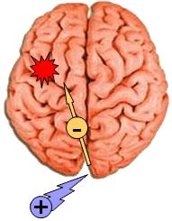
Epilepsy is defined as a “chronic brain disorder characterised by spontaneous recurrent seizures”.The main symptom, seizures, are sudden and temporary events resulting from excessive electrical discharge by neurones. Due to this abnormal electrical activity, the brain waves in epileptic patients are very different from those observed in normal patients.
 |
|
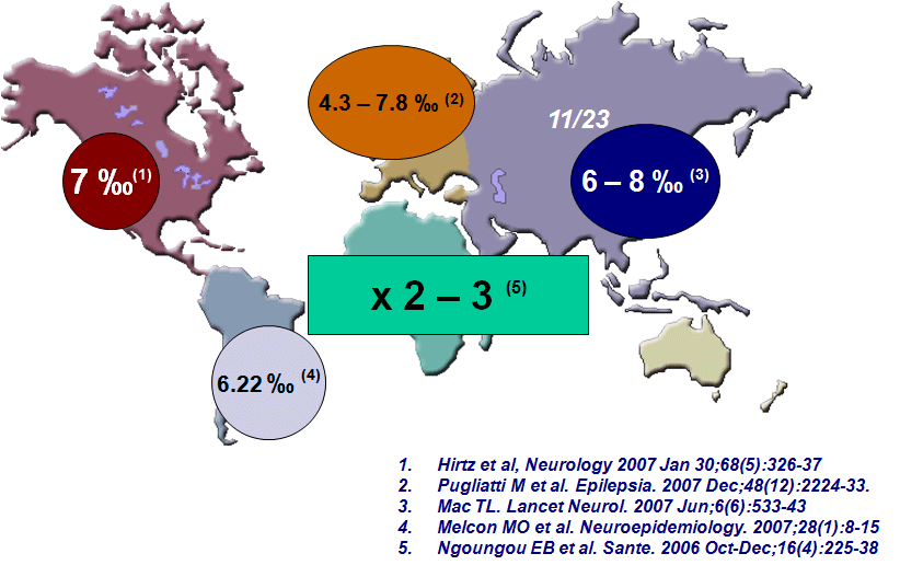
Worldwide incidence of epilepsy
|
TREATMENT OPTIONS
The treatment’s objective is to eliminate or, at least, reduce as much as possible the frequency of epileptic seizures so patients have normal lives as far as possible: children in school and adults at work. If well managed, 75% of patients have their seizures controlled or significantly reduced in frequency.
The large majority of patients have their seizures controlled with anti-epileptic drugs (AED). The main concern with AED is that, though they have different efficacy rates, all share side effects such as sedation, irritability, neurologic problems, reduction in blood cells, or depression. They can even produce behavioural disorders. The patient’s discipline (taking the AED on time, no alcohol or drugs, keeping to schedules: going to bed on time and not staying up late) is, thus, essential to obtain optimum results.
However, some patients are unresponsive to drugs. For those, surgical treatment may be considered. An exhaustive study, carried out by a team of specialists, will be needed, and the neurologist, in one last attempt, will administer new AED to which the patient could respond. Were this case, no additional actions would be needed. However, in most cases this change of AED will not suffice and it will be necessary to continue diagnosis to gather the information needed for the surgical procedure.
Different neuro-imaging techniques will be used, such as CT scan or, more frequently, MRI. The results of these techniques may show the presence a lesion that justifies the epileptic seizures. MRI can occasionally show tumours, vascular malformations, and, in general, lesions that could provoke irritation of nervous tissue and the consequent seizures. High field MRI is especially useful since it can show neuronal migration disorders and so pinpoint the possible areas of seizure onset. Overall, the results of these techniques will have to be correlated with those of EEG and with the patient’s clinical features.
 |
 |
 |
|
CT scan of brain tumour inducing epileptic seizures
|
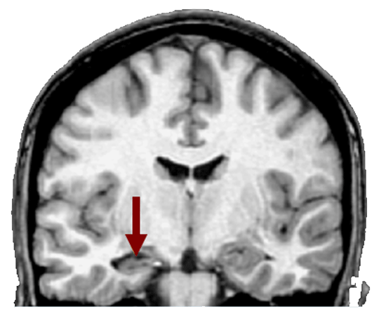
MRI coronal section of mesial temporal lobe slerosis inducing epileptic seizures
|
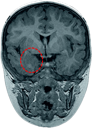
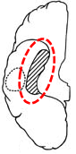
MRI showing cortical dysplasia
|
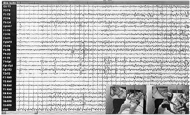
Next, a series of EEG recordings will be gathered to try to pinpoint exactly where the area of seizure onset is and, if it is impossible to achieve an exact diagnosis of its precise origin, the patient will be admitted for a video EEG. This means ALL anti-epileptic medication is withdrawn and continuous EEG recordings are taken for a few days while monitoring the seizure with a video camera to discover the area of onset. Nuclear medicine imaging techniques may be performed during seizures. These techniques may show the seizure origin point using radioactive isotopes and, once more, their results will be compared to those of other techniques to see if they agree on the likely point of seizure onset.
 |
 |
|
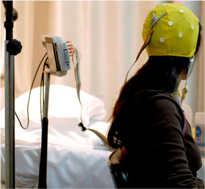
Electrode placement on the scalp for video-EEG
|
Video EEG image
|
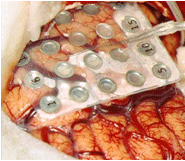
All in all, sometimes the type of epileptic seizures and their exact origin can be diagnosed, others they can’t. In this last case, if the approximate area of onset has been determined, the next step is surgical procedure. Electrodes may be implanted in the brain to record electrical activity as close as possible to the point that is suspected of being the area of onset; other times a subdural grid of electrodes is placed on the brain and are left for a few days to try to obtain more information on the type of seizures and their origin.
 |
 |
|
Subdural grid of electrodes surgically placed on the brain
|
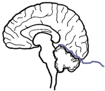
Electrodes on cerebellum to control epileptic seizures
|
 |
With the results of all these techniques, our team will assess your particular case. Depending on the type of seizures, their origin, the damaged area of the brain, the age of the patient, and on many other factors, we will determine the best surgical treatment for you.
|
|
Assessment electrophysiology studies
|
Candidates for surgical treatment of their seizures must, as a general rule:
- be medically intractable
- have a single epileptogenic focus
- have a syndrome which is surgically remediable
- not have degenerative diseases or non-epileptic seizures
If the patient presents a tumour, vascular malformation, etc., that is the cause of the epilepsy, surgical resection of the whole lesion will be the next step to take. Very frequently, when the lesion and the surrounding tissue, damaged by the haemorrhages, are removed, epileptic seizures are eliminated. For the rest of cases, there are three surgical options:
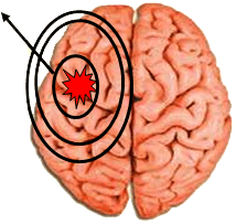
- Removal or elimination of the epileptogenic focus. This is only possible when there is a single focus that is located on expendable areas of the brain, as is the case with temporal lobe seizures and with seizures caused by neuronal migration disorders if the problematic area is surgically accesible and if its removal won’t cause neurological sequelae (loss of the ability to speak, inability to move one or more limbs…).
- Disconnection: destroying the spreading pathways of the epileptic seizures from the point of origin to the rest of the brain. This is achieved by cutting these pathways. A good example is the sectioning of the corpus callosum, which is a great pathway that connects both hemispheres of the brain.
- Stimulating other areas of the brain or of the Peripheral Nerve System (PNS) to reduce the quantity and severity of the seizures.
 |
 |
 |
|
Removal of the epileptogenic focus
|
Sectioning of propagation pathways of epileptic seizures
|
Stimulation of the brain or of the PNS
|
The best results are obtained when it is possible to remove the focus where seizures originate; the rest are palliative treatments with lower effectiveness.
 |
 |
 |
|
Removal of the focus: amygdala-hippocampectomy of the temporal lobe
|
Disconnection: callosotomy
|
Neurostimulation: Vagal stimulator
|
 |
 |
|
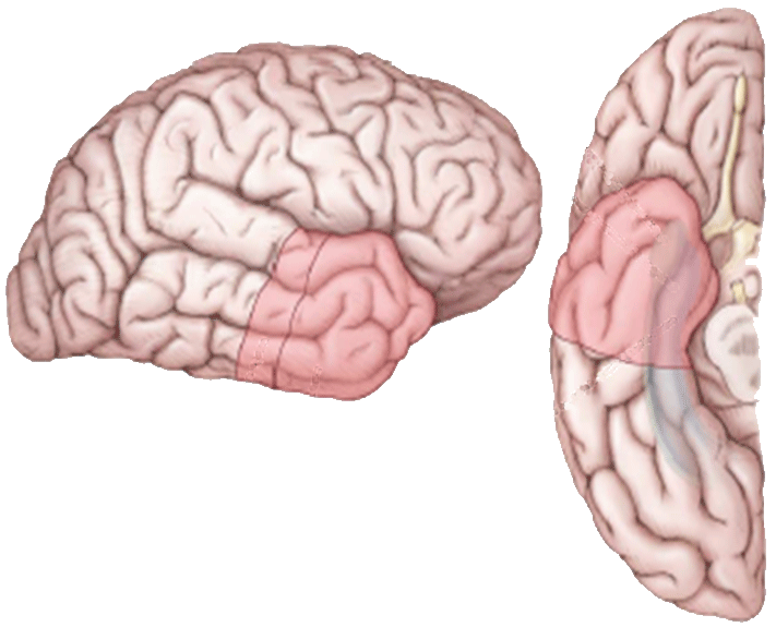
Removal of the focus: lobectomy of the temporal lobe
|
Removal of the focus: functional hemispherectomy (removal of the central area of the hemisphere and disconection of the rest)
|