BRAIN ANEURYSMS
 |
A cerebral aneurysm is a bulging, weakened area in the wall of a cerebral artery, like a very thin balloon or a weakened part of an inner tube. Their main problem is that this weakness in the wall of the artery is susceptible of cerebral haemorrhage. This bleeding, known as subarachnoid haemorrhage, is arterial type and of sufficient severity to that 60% of patients with this type of haemorrhage dies before reaching the hospital. Many aneurysms, fortunately, are diagnosed before they bleed, giving an option to treat them before they cause serious problems.
|
|
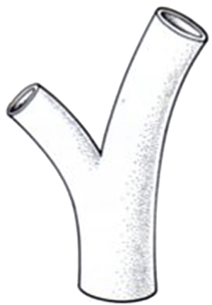
Parts of a cerebral aneurysm
|
 |
 |
|
Normal cerebral artery
|
Saccular brain aneurysm
|
Based on its shape they are classified into saccular and fusiform. Saccular aneurysms are the most common and are like a balloon with a defined neck and a large portion called ‘dome’, which stands only on one side of an artery wall. The fusiform aneurysms are less common and it is an expansion of the entire arterial wall all around it, and therefore has no definite neck. This makes their treatment much more complex.
 |
 |
|
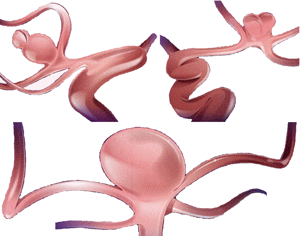
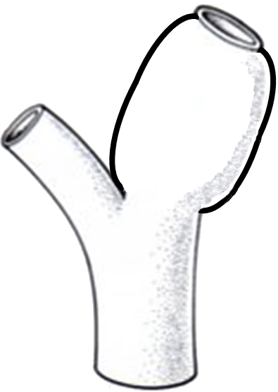
Types of brain saccular aneurysms
|
Fusiform brain aneurysm
|
Why do brain aneurysms develop?
 |
Cerebral aneurysms are caused by a weakness in the artery wall. This weakness may be due to birth defects in the wall of the artery, anatomical variations of the arteries that cause abnormal high blood flow through a given point (usually aneurysms occur at junctions or where they are branches of main trunk), and some are due intake of some toxics, among them the tobacco. Occasionally, they may be due to an inherited disorder of the connective tissues (e.g. polycystic kidney disease or Ellen-Danlos syndrome), an infection or a trauma (for example, a penetrating injury to the brain injuring the wall of an artery).
|
|
Wall of a brain aneurysm
|
Symptoms
Although unruptured cerebral aneurysms may be associated with headaches, in most cases they do not. Any of these symptoms may be present:
- Loss of sensation
- Dilated pupils
- Double vision
- Pain above and behind the eyes
- Headache located in a persistent point
People who suffer a ruptured brain aneurysm (subarachnoid haemorrhage) often have bleeding prior to the warning signs. Warning signs that precede about 40 percent of all large aneurysm ruptures are:
- The worst headache of your life
- Nausea and vomiting
- Stiff neck
- Blurred or double vision
- Sensitivity to light (photophobia)
- Loss of sensation
 |
 |
|
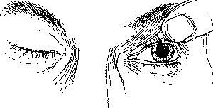
Dilation of the pupil in the case of a cerebral aneurysm
|
Eyelid drop in the case of a cerebral aneurysm
|
What is important about brain aneurysms?
 |
A cerebral aneurysm may rupture, causing significant bleeding (subarachnoid hemorrhage) inside the head. This often produces a sudden and very severe headache (the worst headache of your life!). If bleeding is significant, it is normal for the patient to lose consciousness. In very grave bleeding, there is always the possibility of sudden death.
|
|
Rupture of a brain aneurysm
|
Often, the small orifice of the aneurysm heals, bleeding stops and the person survives. However, the risk of re-bleeding is high, so that the aneurysm should be treated as soon as possible. In severe cases, bleeding may cause brain damage with paralysis or coma. In more severe cases, bleeding can cause death.
 |
 |
|
Image of intra-ventricular hemorrhage with hydrocephalus
|
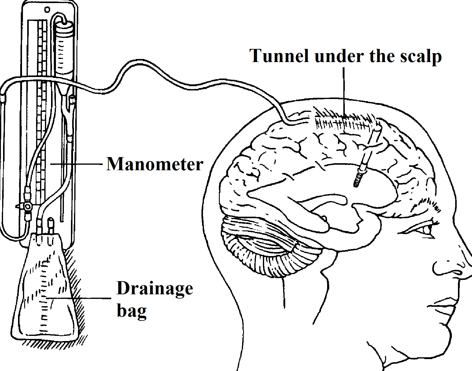
External ventricular drainage
|
Bleeding from a cerebral aneurysm may have several consequences. Blood pools around the base of the brain and blocks the normal flow of cerebrospinal fluid (which is secreted within the brain and has to move until it is absorbed into the existing veins at the top of the brain). This called hydrocephalus. The accumulation of cerebrospinal fluid is treated by inserting a catheter (silicone tubing) inside the brain cavity (ventricle), which secretes the fluid. This silicone catheter drains the bloody fluid into a bag placed next to the patient’s bed (external ventricular drainage).
The blood that accumulates around the base of the brain can also cause a condition called vasospasm. This vasospasm usually occurs typically between 3 and 14 days after the initial haemorrhage. The blood that is out of blood vessels at the base of the brain makes these vessels become irritated and contract. A strong contraction of these blood vessels can cause lack of blood supply to the brain and, in the most extreme cases, even a cerebrovascular accident (stroke). To treat vasospasm, blood pressure is raised through medication. Other drugs may also be given to reduce the threat of vasospasm. Finally, catheters are sometimes introduced into the artery to dilate the narrowed vessels with a balloon, a drug, or both. Vasospasm and the risk of suffering it are reduced with the passage of time.
Ruptured aneurysms are treated in general as soon as possible (one or two days) to prevent new bleeding and to raise the patient’s blood pressure to treat vasospasm.
Diagnosis
Today, thanks to magnetic resonance imaging (MRI) and computed tomography (CT) brain aneurysms can be detected with increasing frequency even before they rupture and bleed. Once a brain aneurysm is detected, it must be treated immediately to prevent further problems. The fact that the aneurysm is not broken is no excuse to unduly delay the treatment.
MRI
 |
 |
|
MRI image of an aneurysm of the basilar artery
|
MRI image of an aneurysm of the basilar artery
|
Magnetic resonance imaging (MRI) is a safe and painless diagnosis which examines different parts of the body through a long magnetic tunnel shaped like a donut and a computer. The magnetic signals from the part of the body examined are displayed using computed radio waves. The computer is able to turn radio waves into images and provide physicians with an image of the aneurysm.
MRA
 |
Magnetic resonance angiography or MRI angiography (MRA) combines MRI imaging with the injection of a contrast dye into a vein. Images are taken after the injection of the contrast agent. A computer is used to reconstruct the 3-D vessels in order to better define the anatomy of the aneurysm.
|
|
Image of magnetic resonance angiography
|
CT scan
 |
 |
|
CT scan image of a subarachnoid haemorrhage
|
CT scan image of a thrombosed basilar artery aneurysm
|
A computed tomography (CT) is a diagnostic method, painless and secure, which examines cross-sections of the brain with a computer and X-rays. The image of the different cuts performed by the device accurately reflects the anatomical structures inside the brain. A CT scan can not only help us to see the aneurysm, but especially the blood of the subarachnoid haemorrhage.
CTA
A CT angiography (CTA) is used to highlight blood vessels inside the head. This test combines a regular CT scan with injection of a contrast dye into a vein. Once the contrast agent reaches the brain, images are created by a CT. It uses a computer to create pictures in 3-D, to better define the aneurysm. In some cases, the CTA can be the ultimate test before treatment.
Angiography (arteriography)
The angiogram is the most complete imaging detection methods.
Before the procedure
If you are hospitalized, in the pre-admission clinic you will carry out a blood test, a physical examination and a medical history. The night before the procedure you should not eat or drink anything after midnight. On the morning of the procedure you must appear in the neuroradiology suite, where a nurse will give you a hospital gown and you will have to remove glasses, contact lenses and dentures. As in an MRI or MRA, before the angiogram you must remove all metal objects or with magnetic band such as watches and hearing aids.
During the procedure
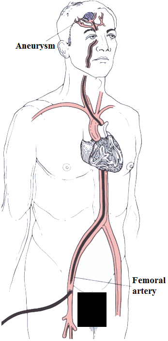 |
Once you are on the stretcher, we will place an intravenous (IV) line on to your arm to provide fluids and medications for your comfort during and after the procedure. It will control your blood pressure and heart rate. The nurse will shave a small area of the groin, where the neuroradiologist will inject a local anaesthetic to numb the area. When the area is anesthetised, the neuroradiologist will insert a catheter into the artery and then inject a contrast agent that will reach the blood vessels of the brain, and X-ray images will then be taken. During the examination you must remain still and follow the instructions of the neuroradiologist. The anaesthesiologist will administer you some drugs to keep you comfortable and help you relax during the study. When the contrast agent is being injected, you will feel warmth in the face and head. Once we have taken all the images, they will be reviewed by the neuroradiologist. If the images are satisfactory, we will remove the catheter from your groin. There are different methods to close the hole in the artery, which may include a locking device or simply to apply pressure on the puncture site for about 15 minutes. Depending on which method is used, you may need to lie down for a short time or for several hours. It is recommended that you relax and avoid strenuous activities for seven to ten days (to prevent bleeding at the puncture point at the groin). |
|
Brain angiography
|
Evaluation
Our team of neuroradiologists, neurologists and neurosurgeons will decide which treatment is best for you.
Options of treatment of the aneurysms
Today, the first treatment to consider is the endovascular, for which a catheter it is inserted through an artery in the groin, allowing the placement inside the aneurysm of a small coil of metal (usually platinum) that causes clotting of blood (thrombosis) inside the aneurysm. This prevents risk of bleeding, at least initially. Not all aneurysms can be treated with this procedure, depending on the shape, size and location of the aneurysm. But for those which can be treated with this procedure, coil embolization allows an effective and minimally invasive treatment. Other devices can be used to assist in coiling, like a balloon or a stent, a metal mesh tube that is placed in the artery.
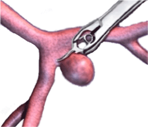
If for any reason endovascular treatment is not possible, surgical proceduresurgical procedure is indicated instead. For the operation a small opening in the skull (craniotomy) is performed and after separation of the brain, the aneurysm is identified. Then a metal clip is placed at the neck of the aneurysm, obliterating it and avoiding the risk or re-bleeding. During the surgical procedure and also during the embolization, the aneurysm may bleed, complicating treatment and reducing the chances of success.
 |
 |
|
Surgical treatment of a cerebral aneurysm with clipping
|
Cerebral aneurysm embolization with coils |
Problems that may arise after the rupture of an aneurysm, such as vasospasm, hydrocephalus, and fever, are usually treated in intensive care unit. In some cases may occur late effects of haemorrhage as headaches, seizures, stroke or hydrocephalus, which may require additional medications or surgical treatment.
Post-operative care
Once the embolization procedure is over, you will undergo a clinical examination. You may be referred to the ICU or recovery room for several hours, as appropriate. The procedure can be performed under general anaesthesia (especially in children), in which case the patient remains hospitalized. In adults the procedure is usually performed under local anaesthesia only, and the patient is awake during the procedure.
You may need to keep your leg straight for a while after the procedure, according to the method by which the neuroradiologist has withdrawn the catheter from the artery.
The time spent in hospital varies. Patients usually return home on the morning after the procedure, but others stay longer in hospital.
Are aneurysms hereditary?
The familial incidence of cerebral aneurysm is rare but can happen. In this case it is recommended that all family members undergo a MRI study, MRA or CTA. In most cases, aneurysms are not hereditary and there is one case in a given family. However, occasionally, a family has two or more members affected by this disease. It has been estimated that the rate of cerebral aneurysms in families with subarachnoid haemorrhage patients is around 10 percent..