HERNIATED CERVICAL DISC
WHAT IS A HERNIATED CERVICAL DISC?
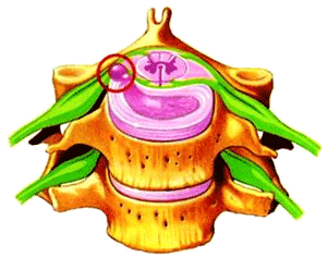 |
Intervertebral discs are the structures between the vertebrae that allow the spinal column to move. When these discs degenerate with age or repeated efforts, their outer layer (annulus) may crack. This means that the disc’s contents may actually slide from their original location, resulting in what is known as a herniated disc. The displaced material may compress nervous structures, such as the spinal cord or its nervous roots. Although there are discs throughout the entire spine, herniated discs appear mainly in the neck and in the low back area low back area. |
|
Cervical disc hernia
|
WHAT IS CERVICAL SPONDYLOSIS?
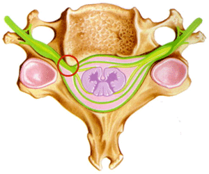 |
Cervical spondylosis is a degeneration of the joints between the cervical vertebrae, including the disc, due to repeated minor trauma, rheumatic diseases or age. This degeneration shows up as ridges, peaks or osteophytes, all of which can behave the same way as a herniated disc, compressing the spinal cord and/or its nerve roots. Herniated discs are softer than osteophytes, but, often, both are found in the same patient. |
|
Hard herniated disc caused by an osteophyte compressing a nerve root
|
WHAT ARE THE SYMTOMS?
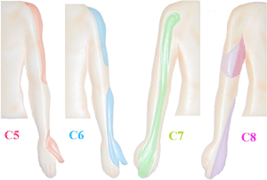 |
Symptoms vary greatly from one patient to another, and may range from minimal discomfort to inability to walk. When herniated cervical discs compress a nerve root, there is typically pain and stiffness in the neck and pain and/or numbness in arms, hands and fingers, as well as dizziness and, occasionally, loss of sensitivity in the arms. It is rare for herniated cervical discs to compress the spinal cord, causing impairments in the lower limbs (which affect the gate) and in sphincter control, which induces difficulties to control urination.
|
|
Areas of pain and sensory loss due to herniated cervical disc
|
 |
 |
|
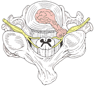
Herniated cervical disc radicular compression and pain in the upper limb
|
Herniated cervical disc with cervical spinal cord compression and paralysis in the lower limbs
|
HOW IS DIAGNOSIS MADE?
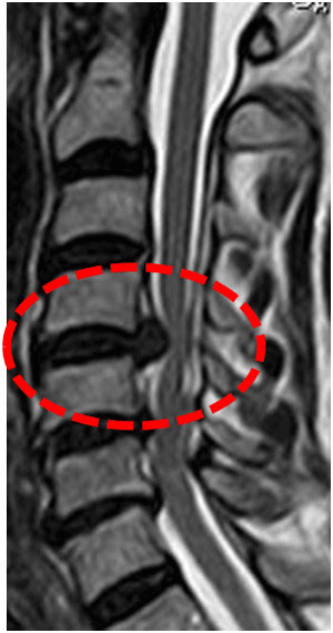 |
Diagnosis is made based on the symptoms and a neurological examination. Plain X-rays and CT scans can show spondylosis, but MRI scans allow a more accurate diagnosis, especially of herniated discs and the structures it has affected. Sometimes an electromyography is used to identify the affected nerve.
|
|
MRI image showing a herniated cervical disc compressing the spinal cord
|
WHEN IS SURGERY NEEDED?
In most cases, medication and rest is the solution. Once the acute phase is over, physiotherapy is recommended in addition to the other conventional treatments. However, if the disc is herniated greatly, if there is damage to nerves, or if conventional therapies fail, then surgical treatment may be required.
WHAT SURGICAL PROCEDURES MAY BE CARRIED OUT TO TREAT A HERNIATED DISC, CERVICAL SPONDYLOSIS, OR BOTH?
The choice of technique and of approach (anterior or posterior) will depend on the case and, sometimes, on the experience of each surgeon.
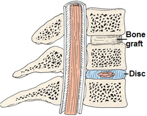
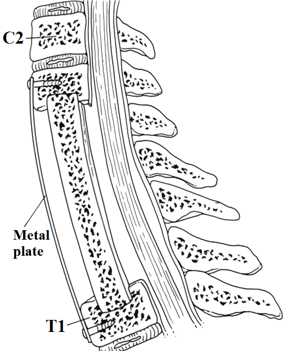
The anterior approach is carried out through the folds of the skin on one side of the neck (either right or left). The aim of the procedure will be to remove the herniated disc and/or osteophytes. After disc removal, often a bone graft, or a graft made from another material, will be inserted to compensate for the lack of stability between the two vertebrae (anterior arthrodesis). Some surgeons prefer to remove almost all of the vertebral body instead (corpectomy) and, after placing a bone graft, to immobilise the vertebrae with titanium plates. In young people with good facet joints, replacing the damaged disc by a disc prosthesis is highly recommended. In older people with osteoarthritis at that level, however, a disc prosthesis usually results in neck pain so it is not suitable for this age group.
 |
 |
|
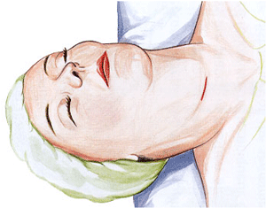
Surgical incision for anterior approach to treat a herniated cervical disc
|
Spinal column exposure during anterior cervical discectomy
|
 |
 |
|
Anterior cervical discectomy to treat herniated cervical disc
|
Bone graft replacing the removed disc
|
 |
 |
|
Multilevel cervical corpectomy
|
Cervical disc prosthesis
|
The posterior approach is carried out through a midline incision in the back of the neck. The back of one or more vertebrae is removed (laminectomy), thus getting rid of the compression on the nerve roots or on the spinal cord. Another option is to use a minimally invasive technique, in which through a 16mm incision the herniated disc is extracted without lesioning the spinous processes or the laminae. On removing the offending disc fragment, the pain in the upper limb usually goes away. However, over time there may be some neck pain.
 |
 |
|
for posterior approach to treat herniated cervical disc
|
Bone resection point for posterior approach to treat herniated cervical disc
|
 |
 |
|
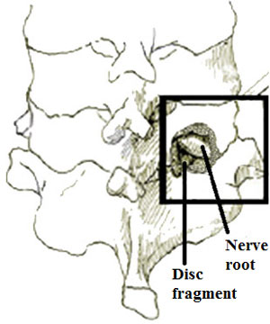
Exposure of herniated cervical disc
|
Removal of herniated cervical disc
|
WHAT ARE THE MOST COMMON AND MOST SEVERE COMPLICATIONS?
The most common complications are neck discomfort and pain when swallowing. They usually subside with time and are well controlled by medication. Occasionally, there may be problems with the bone grafts or disc prostheses placed between the vertebrae, wound infections, and problems derived from general anaesthesia. Though exceptional, these are the most severe complications and happen due to damage to areas close to the cervical spine (spinal cord, oesophagus and trachea), which trigger the consequent paralysis of one or more limbs. Finally, the nerve of a vocal cord may also be damaged, but this is very rare indeed.
POSTOPERATIVE RECOVERY AND TREATMENT AFTER HOSPITAL DISCHARGE
In general, after anterior-approach surgery the patient will be able to get up the next day wearing a cervical collar. The greatest pain will from the graft donor site, the iliac crest, if a bone graft has been used.
With posterior approach the pain is larger due to the muscle damage (inflicted to allow surgical approach), except when a minimally invasive technique is used. In this case, damage to other tissues is minimal and so the pain will be significantly smaller.