SPONDYLOLISTHESIS
WHAT IS SPONDYLOLISTHESIS?
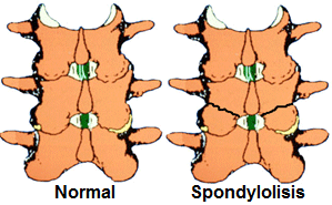 |
About 5% of the adult population suffers from a vertebral fracture at the junction of the facet joints with the pedicle, a site known as isthmus. This is what is known as spondylolysis. It most commonly affects the low back region, just above the sacrum. Due to the constant forces that the area experiences, this fracture cannot heal normally as it would do in other parts of the vertebra or in any other bone.
|
|
Lumbar spondilolysis
|
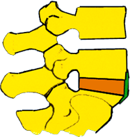
Adult isthmic spondylolysthesis happens when the vertebra with the isthmus fracture slides forward and over the vertebra below. There are other types of spondylolisthesis, though, such as degenerative spondylolisthesis, which is the result of facet joint arthritis and intervertebral disc degeneration.
 |
 |
 |
|
Normal lumbar spinal column
|
Lumbar spondilolysis
|
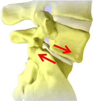
Lumbar spondilolisthesis
|
WHAT ARE THE SYMPTOMS OF SPONDYLOLISTHESIS?
 |
Sometimes, isthmic spondylolysthesis does not cause any symptoms for years. When it does, they may include low back pain spreading to the buttocks, as well as numbness, weakness or pain in the lower limbs (known as sciatica). These symptoms usually worsen when standing and improve with bed rest. Approximately 5-10% of patients with low back pain are suffering from a spondilolysis or spondylolisthesis, but the fact that spondilolysis or spondylolisthesis are seen in lumbar spine X-rays does not necessarily mean that they are causing the symptoms. |
|
Radicular compression due to spondilolisthesis
|
HOW ARE SPONDYLOLISTHESIS DIAGNOSED?
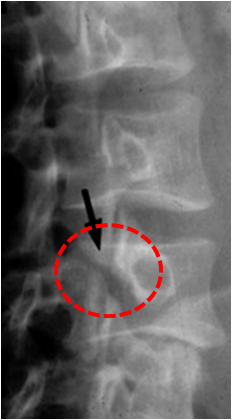
Spondilolisthesis is suspected by the medical history, physical examination and X-rays of the painful area. If X-rays do not show the fracture well, a CT scan may be performed.
MRI can show soft tissues, including intervertebral discs and nerves and their relationship with the isthmic fracture, as well as the degree of sliding of one vertebra over the other. Depending on this degree, spodylolisthesis is graded I (25%), II (50%), III (75%) or IV (100%).
 |
 |
 |
|
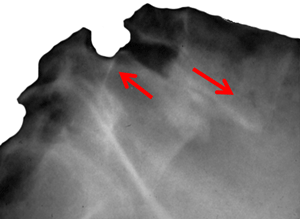
Lumbar spondilolysis
|
Lateral plain X-rays showing lumbar spondilolisthesis |
MR image showing lumbar spondilolisthesis
|
TREATMENT OPTIONS
Conservative treatments will be tried first. These include active rest, i.e. reducing physical activity but trying not to completely eliminate it, as well as anti-inflammatory medication (oral or injectable), analgesics for pain control and physiotherapy to strengthen the muscles of the spinal column.
Medical treatment is usually carried out together with physiotherapy and rehabilitation and involves analgesics, which control pain and inflammation, such as aspirin, ibuprofen or naproxen. They must be taken with a gastric protector to prevent gastritis or stomach bleeding. If non-steroidal anti-inflammatory agents (NSAIDs) do not control the pain, narcotics, such as codeine, may have to be taken for a short period of time. Care should be taken to make sure the dose is the one recommended, for taking a bigger dose will not speed up recovery but will worsen side effects, including nausea, constipation, dizziness and drowsiness, and prolonged use can cause dependence. Due to their powerful anti-inflammatory effects, corticosteroids are sometimes prescribed for low back and severe leg pain, but they can also have side effects such as stomach problems, weight gain, hair growth in abnormal places, and myopathy. They can be taken orally or be injected into the epidural space, around the nerves that branch off the spinal cord, or into the facet joints.
 |
Physiotherapy may include massage, local heat or cold, ultrasound or transcutaneous electrical stimulation. At first, physiotherapy might consist of passive stretches or changes of posture to reduce the low back pain or the symptoms shown by the leg. When the pain subsides, aerobic exercise, such as bike riding or swimming, may be recommended and combined with stretching exercises to improve flexibility and muscle strength in the affected area. Strengthening the abdominal muscles and muscles of the spinal column will help stabilise it. |
|
Orthesis (external brace) to treat low back pain
|
SURGICAL TREATMENT
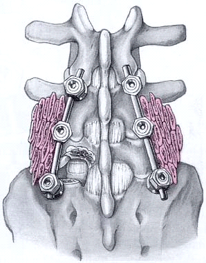
Surgical treatment becomes an option when conservative treatments have failed to control the pain. This pain can be induced by an entrapped nerve, by nearby degenerated intervertebral discs or by the sliding of an unstable fractured vertebra. If a nerve is compressed by the sliding of vertebrae, surgery may be necessary to decompress the nerve. In addition to creating space for the entrapped nerve, the fusion of the vertebrae will be necessary to stabilise the area and avoid future problems; otherwise, the vertebrae may continue to slide and further induce symptoms. To this purpose, a scaffold of titanium screws and bars will be inserted together with bone grafts for the vertebrae to fuse. The surgical approach is usually posterior, but sometimes an anterior approach is necessary to complement it.
Spondilolisthesis surgery success rate is above 75%. After surgery, an orthesis (external brace) will be used for 6 to 12 months, and a post-surgical rehabilitation program may be recommended. IT IS EXTREMELY IMPORTANT NOT TO SMOKE AT ALL, FOR SMOKING PREVENTS BONE CALLUS FORMATION AND, WITH IT, THE DEFINITIVE FUSION OF THE AFFECTED VERTEBRAE.
 |
 |
 |
|
Postero-lateral arthrodesis with a scaffold of screws and bars to treat spondilolisthesis (bone graft in pink)
|
Postero-lateral and interbody arthrodesis
|
Retroperitoneal interbody arthrodesis
|